Medical Mysteries of a Museum Mummy
By Margaret Tamulonis, Manager, Collections & Exhibitions, Fleming Museum of Art
Working in a museum with a mummy in the collection is in some worlds a dream job, and my world is no exception. It was childhood visits to the Metropolitan Museum and the Ancient Egyptian section that launched (or triggered the beginning of) my love of museums and admiration for the people who work in them. When I started at the Fleming Museum at the University of Vermont, it was an additional joy that the Fleming had a real Egyptian mummy—2700 years old, archaeological provenance not entirely clear, purchased for ten dollars by our esteemed original curator in 1910.
The mummy had been on view pretty much since then, and gained pride of place when the new museum building was constructed in 1931. When I’d tell people where I worked, most would comment on the mummy. She is the focus of many school field trips, as well as all sorts of museum tours and certainly pilgrimages by people who grew up in the Burlington area and remembered “our mummy.” For me as a collections manager, she was an object of respect and some unease. It was the first time I had worked in a museum that displayed human remains. At times unknown museum visitors would leave small offerings of corn on top of her exhibition case. Several years into my job, we opened the case and dusted and vacuumed her, to remove an old-fashioned display of sand that had been placed there at an earlier time. But the mummy did not need my regular attention and was safe and sound in the exhibit case (that only needed to be dusted and polished every week for fingerprints).
In 2010, we were approached by an energetic and enthusiastic radiologist from across the street. The Museum shares a driveway with Fletcher Allen Medical Center, Vermont’s large and prestigious medical facility. Jason Johnson, MD (now an Assistant Professor of Radiology and Biomedical Imaging at the University of California, San Francisco and the Chief of Radiology for the 514th Aeromedical Squadron at McGuire Air Force Base), had been researching new methods of CT (computerized tomography) scanning and was intrigued by the use of CT scanning on archaeological materials, including Egyptian mummies. He offered to make the arrangements to scan the Fleming mummy if we were willing, at no cost to the museum.
One of the joys of working at the Fleming is the enthusiasm and support of the staff. Jason first spoke with me (after just asking for me at the front desk), and I passed the request on to our Director Janie Cohen. She was immediately enthusiastic and while we all felt cautious about the process, there was little doubt that we would attempt to scan the mummy if there was no risk of harm.
Dr. Johnson, our then-curator Aimee Marcereau DeGalan, and I met and discussed the logistics, using previous examples of museum mummy scans to set up our process. We knew we would be using an increased radiation dose for the scanner, much more than what would be used on a living being and providing significantly more detail. We knew we would want to do it on a day when both museum and hospital were (hopefully) quiet and with few visitors.
We did know some things about the mummy — she had been purchased in 1910 by the Fleming’s original curator, George Henry Perkins, who had traveled to Egypt. She is from the 25th Dynasty. X-rays performed in 1937 revealed she is female and between 14 and 16 years old. But a CT scan might reveal more, including the possibility of objects wrapped in her linen and some clarification of her cause of death.

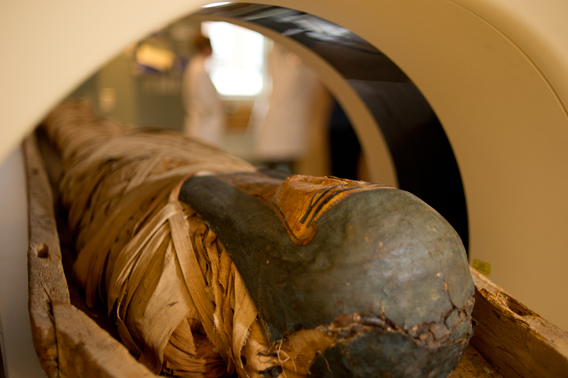
So on a very early and cool November morning, and luckily one without any precipitation, we were able to wheel the mummy on a hospital gurney (carefully covered) the approximately 500 feet from the museum entrance to the closest hospital entrance. A small team of radiologists and a radiology physicist met us at the scanner. Only museum staff handled her and we held our collective breath as we lifted her onto the scanner’s bed, formed for a human and perfect for her and the bottom part of her coffin.
The entire process took two hours, and we were grateful to return to the museum with the mummy after the uneventful but hopefully fruitful scans. And they were fruitful, as we found out a few months later. We convened a group of scientists, academics, and museum staff review the scans, including a UVM anthropologist, the State Medical Examiner Steven Shapiro, and forensic dentist David Averill. The digital scans were also shared with other scientists from further afield, including forensic anthropologists. The conclusions were interesting, and the skull fracture visible on the early X-rays could be viewed in even more detail. While it could still not be concluded that the fracture was the actual cause of death, the images indicated that the fracture had occurred no earlier than a few weeks before her death. In addition, the method for removing the brain in preparation for mummification was through the foramen magnum (the opening in the base of the skull) rather than through the nose or the back of the skull. The scanning methods used for the mummy were also used to develop new standards and methods for forensic scanning.
The scans have continued to be studied for new clues, and these images are a valuable addition to the information we have about the Fleming mummy. Another great result from the multiple scans is a three-dimensional skull that was produced from a 3D printer on the university campus, after we shared the scans with the Instrumentation department. All of this new information and imagery will ultimately be shard in a reinstallation of the Fleming’s Egyptian collection. But the mummy can never be off view for long---she has far too many regular visitors.
Working in a museum means constantly learning new things and meeting new people, and the Fleming Mummy project allowed us to work across departments, professions, and institutions. Luckily, a few of us were just across the street from each other.
Join the author along with Dr. Johnson, Vermont Chief Medical Examiner Dr. Steven Shapiro, Museum Director Janie Cohen, myself, and Dr. Lawrence Berman, Senior Curator of Ancient Egyptian, Nubian, and Near Eastern Art at the Museum of Fine Arts, Boston at the NEMA Conference on Thursday, November 20 , at 11:00 am.